
Study information
Participants in this study received a diagnosis of early-stage or locally advanced breast cancer.
Additional information, including mammograms and imaging reports, as well as biopsy information, was collected.
The study was approved by the Health Research Ethics Board of Alberta - Cancer Committee (HREBA.CC-22-0333).
Sample results
Sample images are shown for a patient with breast density B . This patient was diagnosed with IDC in her left breast, along with extensive DCIS. A localized response is observed in the LMLO view compared to the RMLO view.
Mammograms are shown above the UWB images. The CC views are shown on the left and the MLO views are shown on the right.
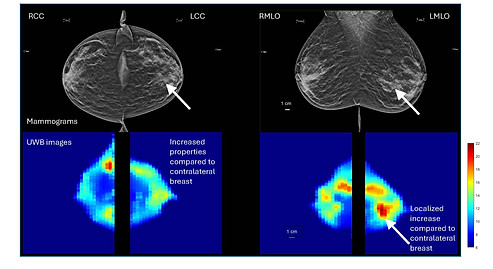
Figure adapted from Mojabi P, Tsang RY, Docktor B, et al. Feasibility of tumor detection with a transmission-based microwave imaging system. Med Phys. 2025; 52:e18080. https://doi.org/10.1002/mp.18080 © 2025 The Author(s). https://creativecommons.org/licenses/by/4.0/. Figures displayed in orientation familiar to radiologists.
Summary results
-
Average properties of UWB images of 13 cancer patients are calculated for CC and MLO views.
-
The ratio of these properties is calculated.
-
Ratios are also calculated for 13 participants with similar ages and breast densities.
-
Ratios are plotted for breast density categories A to D.
-
In this cohort, larger ratios are observed for cancer patients with greater breast densities, particularly in the CC view.

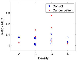
Figure adapted from Mojabi P, Tsang RY, Docktor B, et al. Feasibility of tumor detection with a transmission-based microwave imaging system. Med Phys. 2025; 52:e18080. https://doi.org/10.1002/mp.18080 © 2025 The Author(s). https://creativecommons.org/licenses/by/4.0/. Ratios are plotted for different breast densities.